
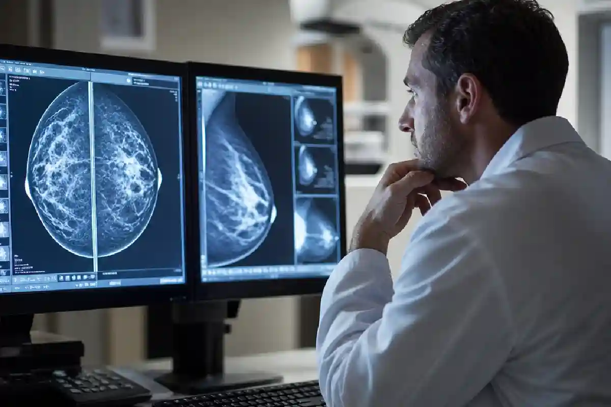



A suspicious finding on a mammogram or breast ultrasound can trigger immediate anxiety, yet most breast abnormalities detected through screening turn out to be benign. The path from detection to diagnosis involves a carefully structured process that balances thoroughness with patient comfort, and understanding what happens between the initial imaging and a definitive answer can help reduce the uncertainty many women experience during this waiting period.
Table of Contents
What Radiologists Look for in Breast Imaging
Breast imaging specialists evaluate several characteristics when reviewing mammograms, ultrasounds, or MRI scans. They assess the shape, margins, density, and distribution of any masses or calcifications. Round masses with smooth borders typically suggest benign conditions such as cysts or fibroadenomas, while irregular shapes with spiculated edges raise concern for malignancy. Calcifications are examined for their pattern—scattered benign calcifications differ markedly from clustered microcalcifications that may warrant tissue sampling.
The Breast Imaging Reporting and Data System, known as BI-RADS, provides a standardised scoring system from 0 to 6. A BI-RADS 3 indicates a probably benign finding with less than 2 per cent chance of cancer, typically requiring short-term follow-up imaging rather than immediate biopsy. BI-RADS 4 and 5 categories indicate suspicious or highly suggestive findings that require tissue diagnosis to rule out malignancy.
The Role of Tissue Sampling in Diagnosis
When imaging results fall into categories requiring tissue confirmation, several biopsy techniques may be employed depending on the location, size, and characteristics of the abnormality. Core needle biopsy has become the standard approach for most palpable and image-detected breast lesions, offering accuracy rates above 95 per cent while avoiding surgical intervention. This outpatient procedure typically uses ultrasound or stereotactic mammography guidance to position the needle precisely within the target tissue.
For women in Abu Dhabi requiring diagnostic procedures, Cleveland Clinic Abu Dhabi’s Breast Health & Cancer Program offers image-guided diagnostic services including breast biopsy performed by radiologists specialising in breast imaging. These procedures collect multiple tissue cores in a single session, providing pathologists with sufficient material to determine whether cells are benign, pre-cancerous, or malignant, as well as specific molecular characteristics that guide treatment planning.
The vacuum-assisted technique allows removal of larger tissue volumes through a single insertion point, making it particularly useful for sampling calcifications or removing small benign lesions entirely. Most patients experience only local discomfort during the procedure and can resume normal activities within a day, though strenuous exercise should be avoided for several days to prevent bleeding or bruising at the biopsy site.
Interpreting Biopsy Results and Next Steps
Pathology results typically arrive within three to five working days, though complex cases requiring additional testing may take longer. Benign results such as fibroadenoma, fibrocystic changes, or fat necrosis generally require only routine follow-up imaging according to standard screening guidelines. High-risk lesions including atypical ductal hyperplasia or lobular carcinoma in situ often prompt recommendations for surgical excision, as these conditions carry increased future cancer risk and may coexist with undetected malignancy nearby.
A cancer diagnosis launches a comprehensive evaluation process. Pathologists determine the tumour type, hormone receptor status, HER2 status, and proliferation rate—all factors that influence treatment recommendations. Additional imaging may be ordered to assess disease extent, and patients typically meet with a multidisciplinary team including surgical oncologists, medical oncologists, and radiation oncologists to develop an individualised treatment plan.
Treatment Pathways Based on Pathology
Early-stage breast cancer confined to the breast and nearby lymph nodes accounts for the majority of diagnoses through screening programmes. Treatment typically begins with surgery—either breast-conserving lumpectomy followed by radiation therapy, or mastectomy with or without reconstruction. The surgical approach depends on tumour size relative to breast size, tumour location, patient preference, and genetic factors such as BRCA mutations that increase risk of additional cancers.
Systemic therapies including chemotherapy, hormone therapy, and targeted treatments are selected based on tumour biology and stage. Hormone receptor-positive cancers respond to endocrine therapies that block oestrogen effects, while HER2-positive tumours benefit from targeted antibodies that inhibit this growth pathway. Triple-negative breast cancers, lacking hormone receptors and HER2 expression, typically require chemotherapy as the primary systemic treatment.
Supporting Quality of Life During and After Treatment
Cancer treatment brings physical and emotional challenges that extend beyond tumour eradication. Side effects vary with treatment type but may include fatigue, nausea, peripheral neuropathy from chemotherapy, skin changes from radiation, and surgical pain or lymphedema. Comprehensive breast cancer care now routinely incorporates supportive care services alongside disease-directed treatment, addressing symptom management, nutritional support, and psychosocial needs throughout the treatment continuum.
Lymphedema prevention has become a focus area following axillary surgery or radiation. Women who undergo lymph node removal receive education about arm protection, exercise guidelines, and early warning signs of swelling. When lymphedema develops, certified therapists provide manual drainage techniques, compression garments, and exercises that help manage this chronic condition.
Surveillance and Long-Term Follow-Up
Completing primary treatment marks the beginning of survivorship care, which includes regular monitoring for recurrence and management of treatment-related effects. Most oncologists recommend clinical examinations every three to six months for the first three years, then annually thereafter. Annual mammography continues for the treated breast and the contralateral breast, as women with breast cancer history face elevated risk of developing a second primary tumour.
Patients taking hormone therapy for five to ten years require monitoring for medication side effects, including bone density loss with aromatase inhibitors and endometrial changes with tamoxifen. Genetic testing results may influence surveillance strategies, with BRCA mutation carriers often advised to consider risk-reducing surgery for the contralateral breast and ovaries after completing childbearing.
The transition from active treatment to survivorship can feel abrupt for some women, who move from frequent medical appointments to less intensive monitoring. Survivorship care plans now document treatment received, potential late effects to watch for, and recommended follow-up schedules, providing a roadmap for patients and their primary care physicians to coordinate long-term health maintenance.
Early detection through screening, accurate diagnosis via appropriate biopsy techniques, and personalised treatment selection based on tumour characteristics have collectively improved breast cancer outcomes substantially over recent decades. Women who understand each step of this process are better positioned to participate actively in decisions about their care and to advocate for comprehensive services that address both disease treatment and quality of life preservation.
Saba Mirza
Saba Mirza is a Health & Wellness Content Writer at Super Healthiness with more than 3 years of content writing experience. She creates clear, accessible and reader-focused content covering health, wellness, fitness, nutrition, personal care and healthy lifestyle topics.
View Author Profile
Sameena Begum
Sameena Begum is a Health, Wellness & Lifestyle Content Reviewer at Super Healthiness with 8 years of content and editorial experience. She reviews selected articles for clarity, structure, consistency, appropriate context and overall reader usefulness.
View Reviewer Profile
Dr. Syed Imtiaz
Dr. Syed Imtiaz is a qualified medical professional with MBBS and MD qualifications and 5+ years of experience in the medical field. He works as a General Physician and Dermatologist and serves as a Medical Reviewer at Super Healthiness, reviewing health-related content for medical accuracy, appropriate context, clarity and responsible presentation.
View Medical Reviewer Profile